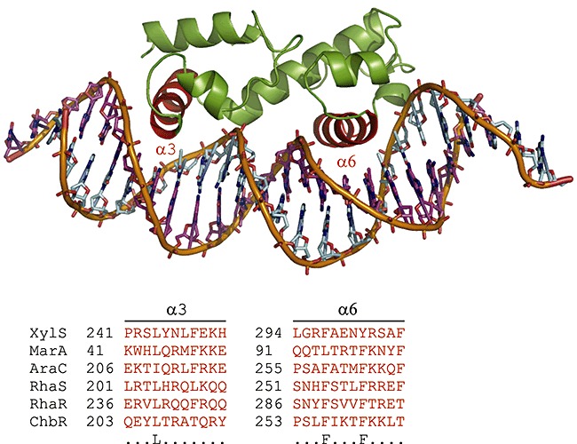
Figure 2.

AraC‐XylS family regulators' characteristic HTH DNA binding motif is shown by using the member MarA as a model (α‐helixes 3 and 6 in red colour). Conserved amino acid residues are depicted in the bottom row. The alignment was derived from the full‐length primary sequences of the given TFs by using the PROMALS3D web server (Pei et al., 2008). Parameters were left at default values. The figure was prepared by using PyMOL (DeLano, 2003). Note that MarA binds DNA as a monomer.
