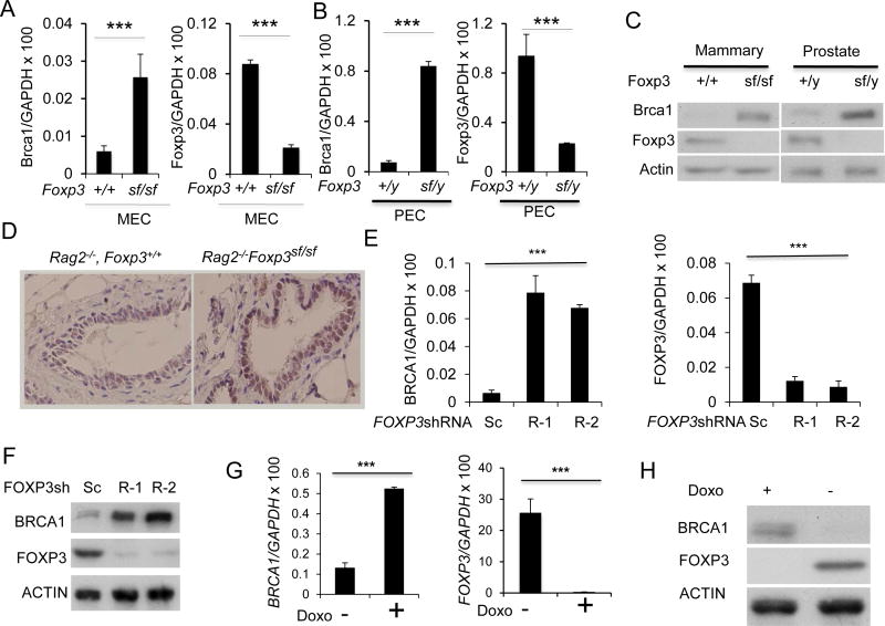
Figure 3.
Foxp3 repressed Brca1 expression in mammary and prostate epithelial cells in the mice. A. Mouse mammary epithelial cells (MEC) were isolated from Rag2−/− Foxp3+/+ and Rag2−/− FoxP3sf/sf mammary glands. Brca1(left panel) and Foxp3 (right panel) mRNA levels were determined by real-time RT-PCR. Data shown were means+/−S.D. of mRNA, presented as %GAPDH and have been repeated three times. ***, P< 0.001. B. Mouse prostate epithelial cells (PEC) were isolated from Rag2−/− Foxp3+/y and Rag2−/− Foxp3sf/y prostates. Brca1(left panel) and Foxp3 (right panel) mRNA levels were determined by real-time RT-PCR. Data shown were means+/−S.D. of mRNA, presented as % Gapdh. The results have been repeated three times. C. The left panel shows Brca1 and Foxp3 expression in MEC by Western blot, while the right panel shows those in PEC. D. Immunohistochemical analysis for Brca1protein in benign mammary tissue from WT and Foxp3sf/sf mice. E–H. FOXP3 represses the BRCA1 gene in normal and malignant human mammary epithelial cells. E. Silencing FOXP3 in MCF10A cells up-regulates BRCA1 mRNA level. The levels of BRCA1 mRNA (left panel) and FOXP3 (right panel) in two scrambled or two FOXP3 siRNA transfected cells as determined by real-time RT-PCR. Data shown are means+/−S.D. of three independent experiments. F. Under the same conditions as in E, MCF10A cells were lysed for Western blot to detect BRCA1 and FOXP3 protein expression as indicated. G. Reduction of BRCA1 mRNA by FOXP3 in MCF-7 Tet-off expression system. MCF-7 cells with Tet-off expression of either GFP control or GFP in conjunction with FOXP3 were cultured for 48 hours in the presence or absence of doxycyclin. The BRCA1 mRNA was quantified by real-time PCR(left panel). The induction of FOXP3 mRNA was shown in the right panel. Data shown are means+/−S.D. of three independent experiments. ***: p value< 0.001. H. BRCA1 and FOXP3 protein were detected by Western blot as shown.

