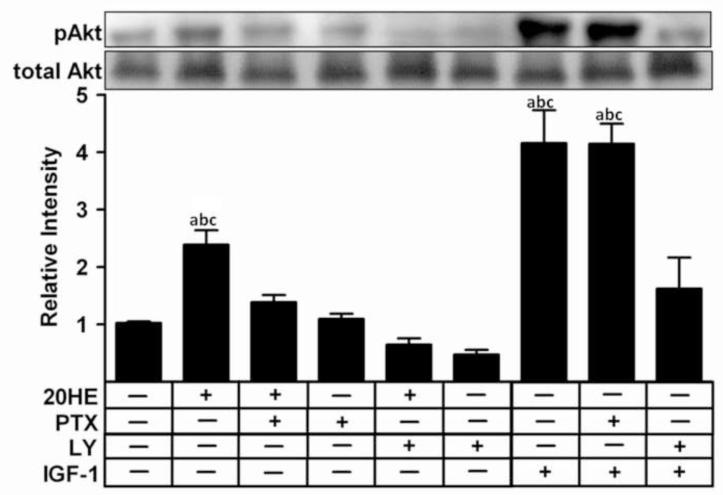
Fig. 4. Effect of G-protein and PI3K inhibitors on Akt phosphorylation in C2C12 myotubes treated with 20-hydroxyecdysone (20HE).
Differentiated myotubes were pretreated for 1 h with either 1 μg/ml PTX, 10 μM LY, or vehicle prior to treatment with either 1 μM 20HE or 100 ng/ml IGF-1 for 2 h. Cell lysates were subjected to Western blotting and probed with either anti-pAkt or anti-Akt. Data shown represent the mean values of phosphorylated Akt normalized to total Akt ± S.E.M. of three experiments. a indicates p < 0.05 compared with control, b indicates p < 0.05 compared with PTX treatment alone and c indicates p < 0.05 compared with LY treatment alone (One way ANOVA/Bonferroni).

