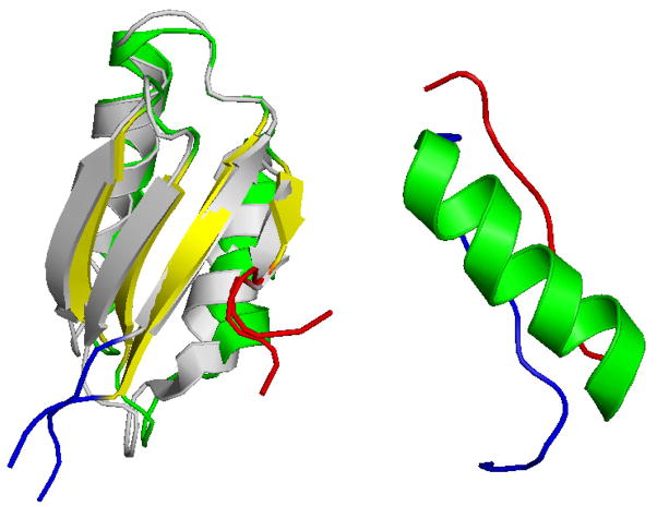
Figure 1.
(Left: Overlay of the wild type (online color yellow/green) and mutant (online color grey) NMR structure of 75-residue MNK6 (PDB-identifier 1YJV and 1YJT) ; right:) NMR structure of the 36-residue protein DS119 (PDB-identifier 2KI0). The online color of the N-terminals is blue, and that of the C-terminals red.

