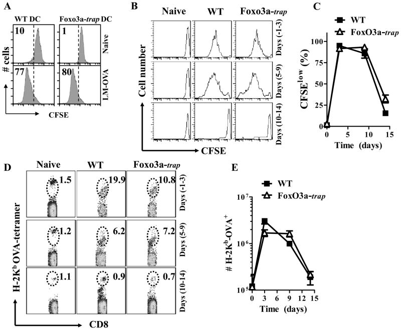
Fig. 3. Similar in vivo antigen-presentation in WT and FoxO3a-deficient mice.
BMDC (5×10⁴) from WT and FoxO3a-deficient mice were infected with 20 MOI of LM-OVA for 30 min. or left uninfected. Extracellular bacteria were removed after washing and incubation in medium containing gentamicin (50 μg/ml). At the end of 2 h, cells were cultured in media containing lower levels of gentamicin (10 μg/ml) and incubated with CFSE labelled OT-1 TCR transgenic cells (106/well). After 3 days of culture, cells were harvested, stained with anti-CD8 antibody, and the reduction in CFSE intensity of OT-1 CD8+ T cells was evaluated by flow cytometry (A). WT and FoxO3a-deficient mice were infected (iv) with 104 LM-OVA. At various time intervals CFSE-labelled OT-1 CD8+ T cells were injected (5×10⁶, iv). Four days after the transfer of OT-1 cells, spleens were isolated from recipient mice and spleen cells were stained with OVA-tetramer and anti-CD8 antibody. Reduction of CFSE expression (B, C) and increase in the numbers of transferred OT-1 cells (D, E) was evaluated. Numbers in the panels represent percentage of OVA-tetramer+ cells within the gate. Results are representative of two independent experiments with three mice per group.

