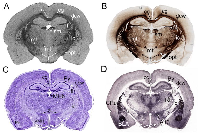
Fig. 1.
Comparison of myelinated structures with several well-defined nuclei on the same coronal plane of 4 different stains. A) On the Gallyas myelin-stained section, it is easy to recognize the cc, cg, ml, mt, sm, f, fi, opt, dcw. B) These same structures can be labeled on the MR image, which is has nearly the same contrast as the myelin stained section. C) On the Nissl-stained section, cellular structures are more evident, such as the pyramidal layer of the hippocampus (Py) and the ventromedial hypothalamus (VMH). D) The acetylcholine esterase stained section reveals different structures again, including caudate putamen (CPu) or the basolateral amygdala (BLA). Taken together, it is evident that a comprehensive, updated myelin atlas of the rat brain is needed that focuses on the white matter structures recognizable on MR images.

