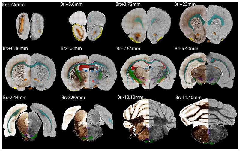
Fig. 2.
Representative coronal sections of the delineated and segmented structures through the brain. This figure shows 12 out of the 120 delineated sections, approximately 2 mm apart from each other. We matched the silver-stained sections (left hemisphere) with the MR images (right hemisphere) and labeled the segmented structures. The color-coded list of the structures that we labeled and analyzed is presented in Table 2.

