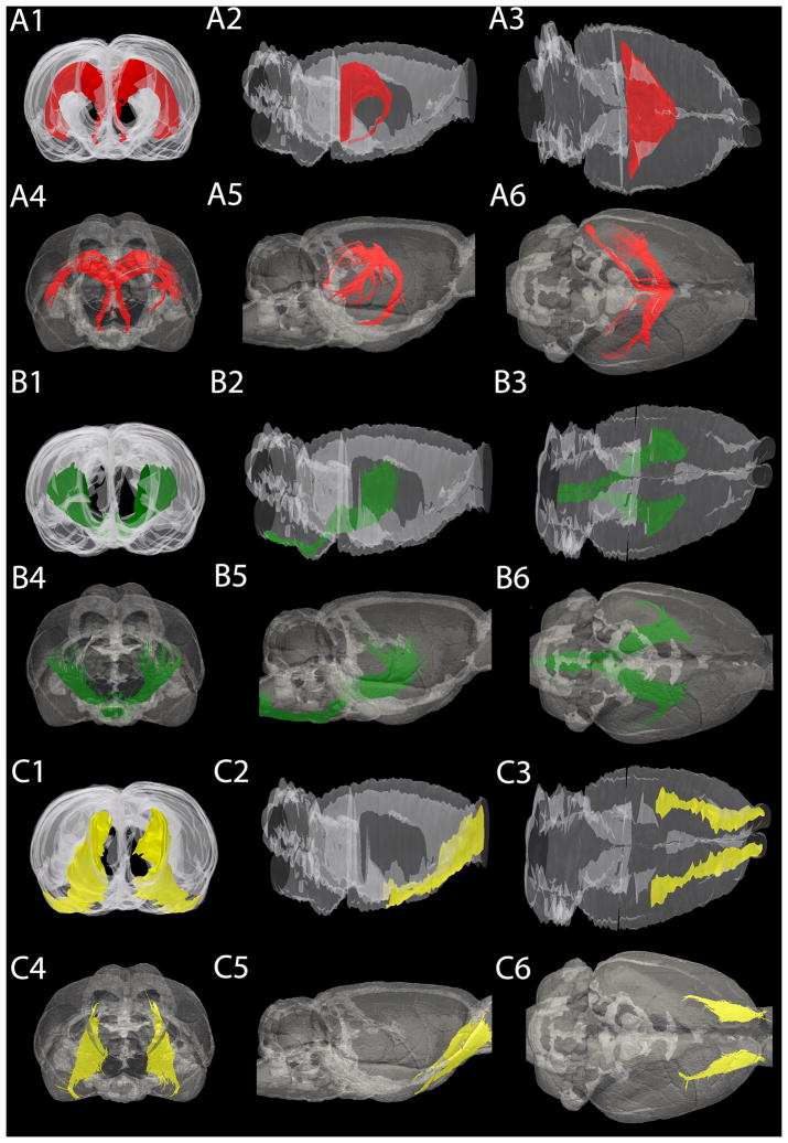
Fig. 3.
Three-dimensional rendering of the rat brain, showing three different structures. (A) Frontal (A1 A4), lateral (A2, A5) and dorsal (A3, A6) view of the fornix/fimbria of the hippocampus/ventral hippocampal commissure. These structures were found to be significantly different based on their volume calculations however; the location and the orientation of the fibers are in agreement. (B) Frontal (B1, B4), lateral (B2, B5) and dorsal (B3, B6) view of the internal capsule. The internal capsule volumes are quite similar between the reconstructed histology and the MR segmentation, and the position and the expansion of the structure shows sufficient overlap. (C) Frontal (C1, C4), lateral (C2, C5) and dorsal (C3, C6) view of the lateral olfactory tract. The lateral olfactory tract shows high inter-modality similarities in terms of volume and spatial extent. In all cases, the outline of the entire brain is shown in transparency for spatial reference.

