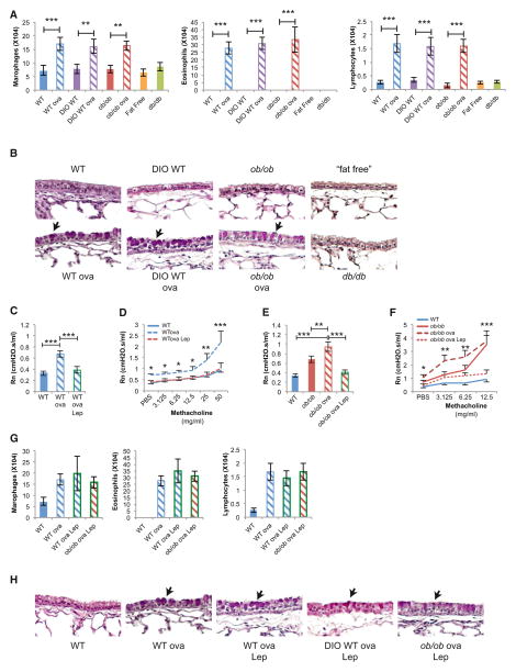
Figure 6. Leptin Regulation of Bronchial Diameter Occurs Independently of Local Inflammation.
(A) Bronchoalveolar lavage analysis demonstrates no difference in inflammatory cells in DIO-WT, ob/ob, “fat-free,” or db/db compared to the ovalbumin (ova)-sensitized and challenged mice (n = 8 per group).
(B) Periodic acid-Shiff staining fails to detect goblet cell (arrows) hyperplasia in DIO-WT, ob/ob, ‘fat-free,” or db/db mice compared to ova mice.
(C–F) Leptin (Lep) ICV infusion in WTova and ob/obova mice decreased Rn and AHR compared to control (n = 12 per group, D-*WTova versus WTova Lep, F-*ob/obova versus ob/obova Lep).
(G) Leptin ICV infusion failed to change the levels of inflammatory cells in WTova, DIO-WTova, ob/obova mice (n = 8 per group).
(H) Leptin ICV infusion failed to change the goblet cell (arrows) hyperplasia in WTova, DIO-WTova, ob/obova mice.
For all panels, *p < 0.05, **p < 0.01, and ***p < 0.001. Error bars represent the SEM. See also Figure S5.

