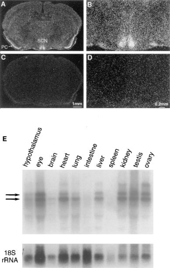
Figure 6. Analysis of Clock mRNA Expression.
(A–D) In situ hybridization of Clock mRNA in the mouse brain. Coronal sections from brains of wild-type mice collected at ZT6, probed with yz50 antisense (A and B) and sense (C and D) ribo-probes.
(E) Tissue distribution blot. Clock mRNA expression is not limited to the SCN. Northern blot analysis of total RNA extracted from diverse tissues (all of which were collected at ZT6 from wild type C57BL/6J mice) reveals that Clock expression is widespread. The bottom panel shows the same filter hybridized with a 32P-labeled oligonucleotide of 18S RNA for normalization.

