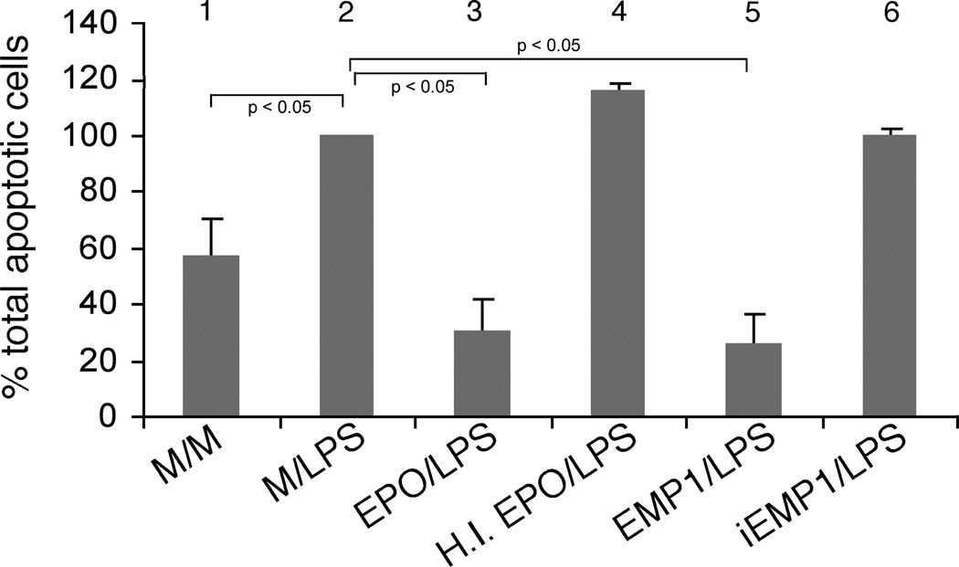
Figure 5.
(A) Expression of EPOR in HUVECs treated with medium alone or EPO (5U/ml) for 24 hours as determined by Western blotting. K-562 lysate was used as a positive control. Data shown is representative of three independent experiments. (B) Expression of EPOR protein in murine aorta. Aortas were harvested from 10 individual mice and whole aortic lysate from each animal was subjected to Western blotting. These data depict 5 of the total of 10 aortic lysates analyzed in this experiment. (C) Expression of EPOR in murine aorta measured by qRT-PCR. These data represent mean ± s.e. of a total of 10 mice. (D) Expression of total Jak2, Stat5 and their phosphorylated forms, respectively, in HUVECs treated with either Medium or EPO for 24 hours as determined by Western blotting. Total HeLa cell extracts (prepared with our without interferon-α treatment and purchased from the manufacturer of the Jak 2 and Stat5 antibodies) were used as positive or negative controls.

