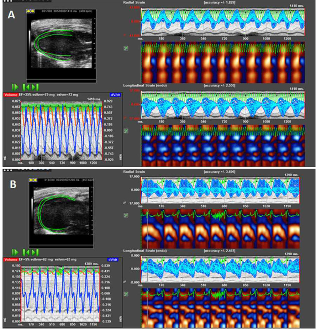
Figure 1.
A representative speckle tracking-based strain echocardiographic analysis of the left ventricle (LV) pre- and post-myocardial infarction (MI). A: Baseline and B: Day 7 post-MI. The post-MI image illustrates LV dilation, reduced ejection faction, and decreased radial and longitudinal strains. Images were acquired with a Vevo 2100 (Visualsonics; our own unpublished data). Analysis was conducted using the VevoStrain™ software.

