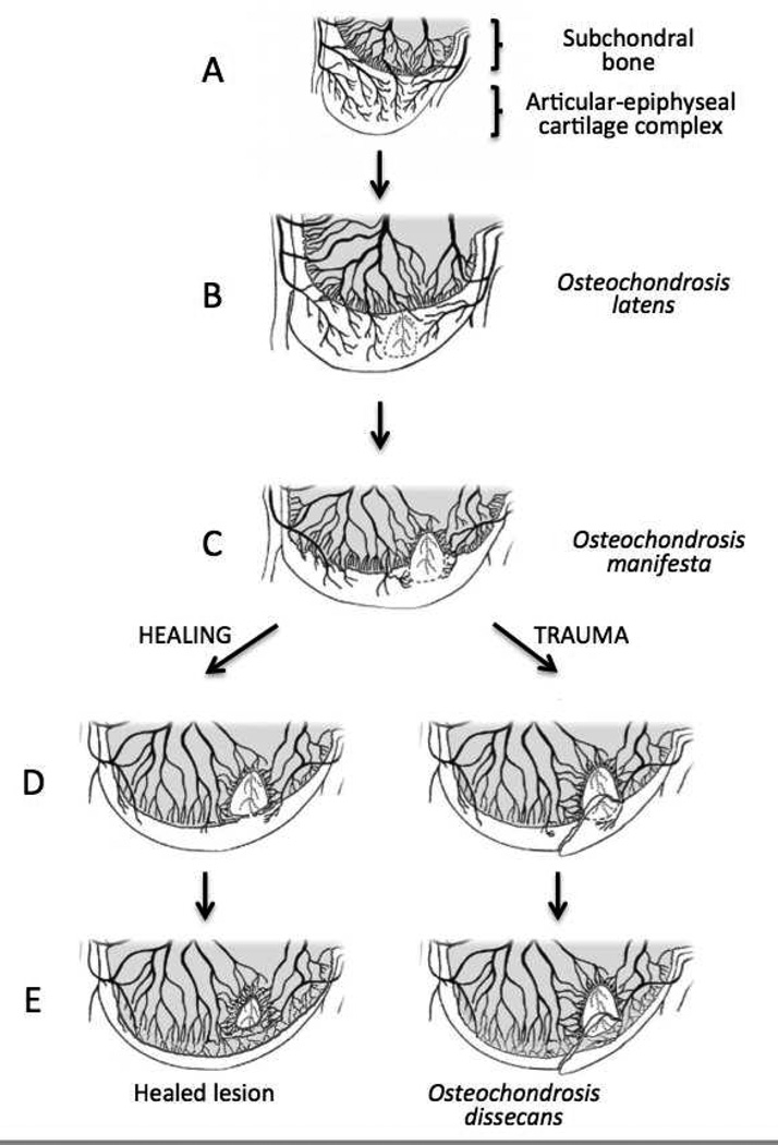
Figure 5.
Diagram demonstrating the pathogenesis of osteochondrosis dissecans (modified with permission from Figure 7,[2]). Panel A: normal enchondral ossification. Panel B: Development of osteochondrosis latens lesion due to failure of cartilage canal blood supply causing necrosis of the epiphyseal cartilage (circled area). Panel C: Osteochondrosis manifesta lesion appears as a delay in the progression of the ossification front. Panels D and E: healing of osteochondrosis manifesta lesion by incorporation into the subchondral bone. Panels F and G: Development of osteochondrosis dissecans lesion due to trauma causing collapse of the articular cartilage overlying areas of necrotic epiphyseal cartilage.

