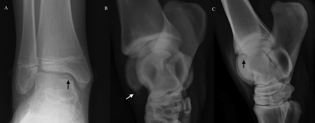
Figure 6.
Osteochondrosis dissecans lesion involving the ankle (tibiotarsal joint). Panel A: posterio-anterior radiographic image of an osteochondrosis dissecans lesion (black arrow) of the talus in a juvenile human subject. Panel B: dorsomedial-plantarolateral oblique radiographic image of an osteochondrosis dissecans lesion (white arrow) involving the lateral trochlear ridge in a horse. Panel C: lateromedial radiographic image of an Ooteochondrosis dissecans lesion (black arrow) involving the distal intermediate ridge of the tibia in a horse.

