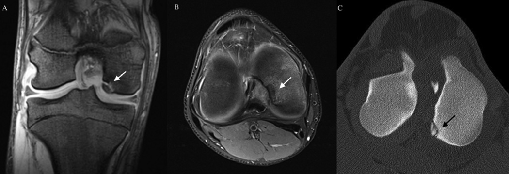
Figure 7.
Osteochondrosis dissecans lesion of the medial femoral condyle. Panels A (coronal plane) and B (transverse plane) depict MRI findings from a human subject with osteochondrosis dissecans of the medial femoral condyle (white arrows). Panel C: CT image of an osteochondrosis dissecans lesion (black arrow) of the medial femoral condyle of a horse obtained in the transverse plain. (Image is courtesy of Dr. Bergman, VetCT-Lingehoeve Diergeneeskunde, Netherlands)

