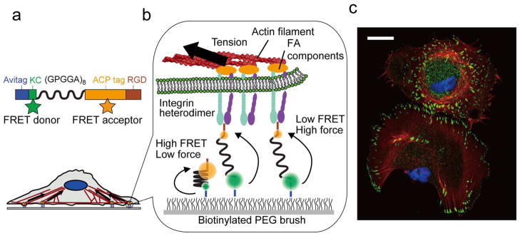
Figure 1.

MTS overview. (a) Sensors are site-specifically labeled with biotin (Avitag), the FRET donor Alexa 546 (KC), and FRET acceptor CoA 647 (ACP tag) and present the RGD sequence from fibronectin (TVYAVTGRGDSPASSAA). The (GPGGA)8 sequence acts as an entropic spring that is stretched upon application of force. (b) Sensor molecules are attached to a coverslip via biotin and Neutravidin; the biotinylated PEG brush prevents nonspecific cell and sensor attachment. Integrin heterodimers attach to the RGD domain and apply load generated by the cell cytoskeleton. (c) Immunofluorescence image of fixed human foreskin fibroblast cells seeded on a MTS-functionalized surface; note the prominent actin stress fibers and FAs (blue: nucleus; red: actin; green: paxillin). Scale bar: 25 μm.
