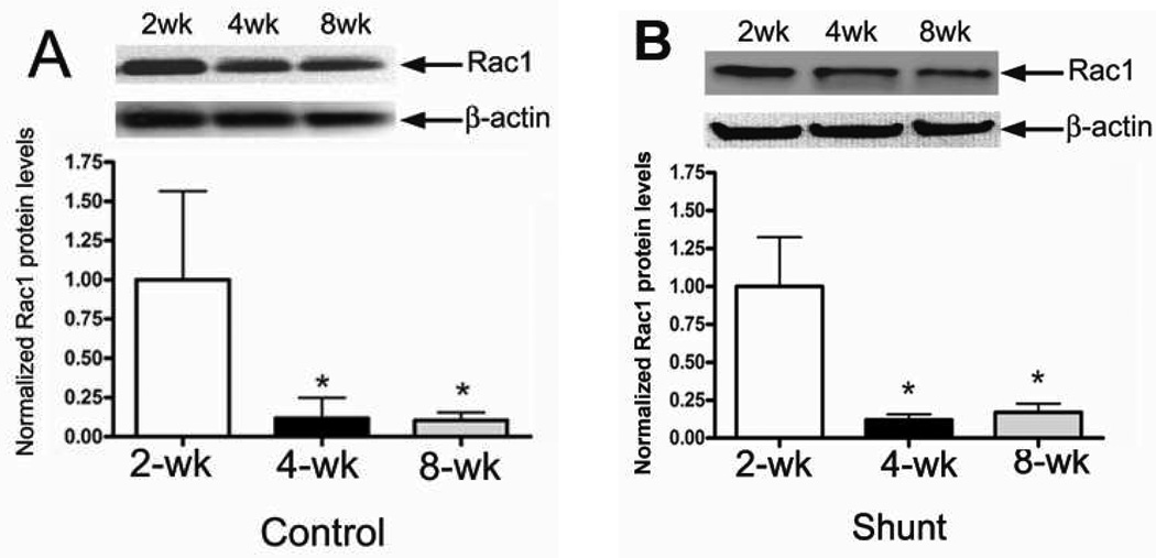
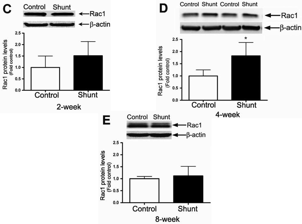
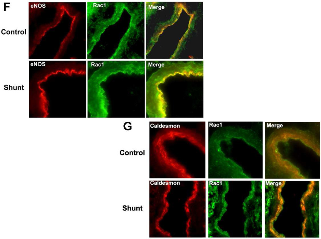
Figure 3. Developmental changes in Rac1 levels in peripheral lung in Control and Shunt lambs at 2-, 4-, and 8-weeks of age.
Protein extracts (50 µg), prepared from peripheral lung of Control (A) and Shunt (B) lambs at 2-, 4-, and 8-weeks of age, were separated on a 4–20% Tris-SDS-HEPES gel, electrophoretically transferred to a PVDF membrane and analyzed using a specific antiserum raised against Rac1. Rac1 protein levels were normalized for loading using β-actin. A representative blot is shown in each panel. Developmentally there was a significant decrease in Rac1 protein levels in both Control (A) and Shunt (B) lambs at 4- and 8-weeks compared to 2-weeks of age. Values are mean ± SD; n=6 control and n=6 shunt at each age. *P<0.05 vs. 2-weeks of age. The protein extracts (50µg), prepared from peripheral lung of 2-(C), 4-(D), and 8-week (E) Control and Shunt animals, were also analyzed to determine changes in Rac1 protein levels between Control and Shunt lambs. Again Rac1 protein levels were normalized for loading using β-actin. Each panel contains a representative blot. There was a significant increase in Rac1 protein levels in Shunt lambs at 4-weeks but no changes at 2-or 8-weeks of age. Values are mean ± SD; n=6 control and n=6 shunt at each age; *P <0.05 vs. control. In vivo localization of Rac1 protein in pulmonary vasculature of 4-week old Control and Shunt lung tissues is shown (F–G). eNOS was used as an endothelial cell marker and Caldesmon as marker of smooth muscle cells. A stack of images was taken every 2µm using confocal microscopy to identify actual co-localization. Magnification is ×20.


