
Figure 2.
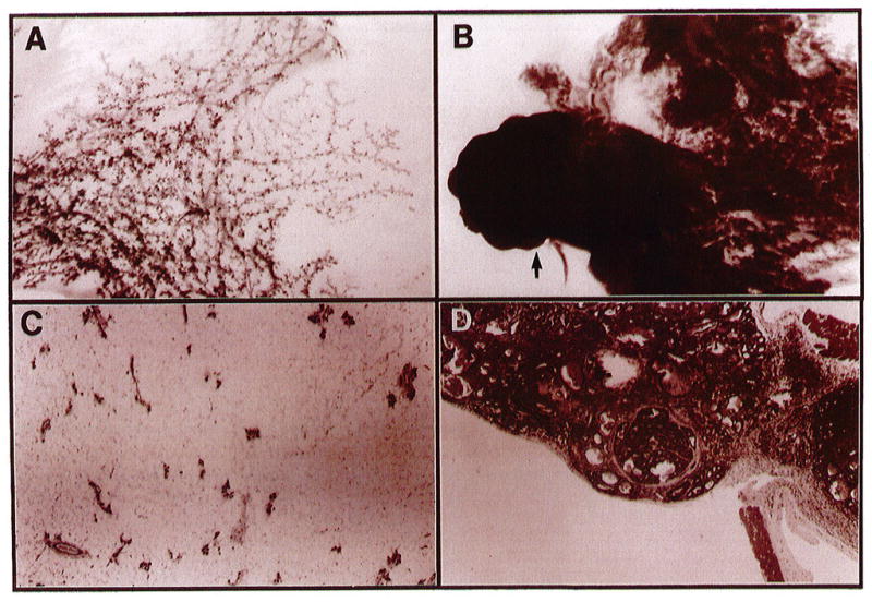
Morphology of mammary glands from normal and SL-1-overexpressing transgenic mice. Whole-mount carmine-stained glands from normal (A) and transgenic tumor-bearing (B) mice and the corresponding hematoxylin-eosin-stained cross sections, C and D, respectively. The arrow in panel B corresponds to the enlarged cross-section in panel D, which demonstrates an encapsulated adenocarcinoma. Both glands are the thoracic (nos 2/3) taken 4 months after the first lactation. Photographs in A and B are 6 × magnification and C and D are 40 × magnification.