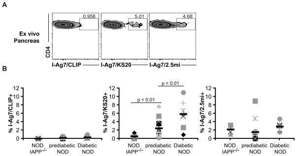
FIGURE 2. Ex vivo analysis of pancreatic infiltrate in NOD mice.
Single cell suspensions were prepared from pancreas of non-diabetic female NOD mice (n = 17; age 10–28 weeks), female NOD.IAPP−/− (n = 4; age 18 weeks) and male and female diabetic NOD mice (n = 7; age 17–26 weeks). Cells were then stained with I-Ag7/CLIP, I-Ag7/KS20 or I-Ag7/2.5mi tetramers. After 2.5 h, cells were stained with a master mix of antibodies and gates were set on live/singlets/CD4+ CD45+ TCRβ+/CD8− CD11b− CD11c− F4/80− CD19− 7AAD− cells. A. Tetramer staining from the pancreas of one representative prediabetic NOD mouse. B. Scatter plots show the percentage of tetramer-positive cells in the pancreatic infiltrate for each tetramer. Each symbol represents an individual mouse and black dashes represent averages. Data are compiled from 2 independent experiments for NOD.IAPP−/− and diabetic NOD and 4 independent experiments for non-diabetic NOD.

