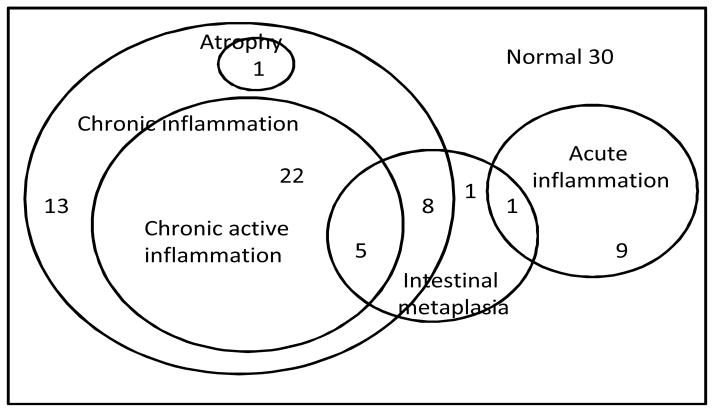
Figure 1.
Venn diagram of overlapping histological abnormalities in 90 controls. A majority of the controls had chronic inflammation, much of which was active, and a minority of which was associated with intestinal metaplasia. Histological evidence of atrophy was seen in only one control, though serological markers suggested 17% prevalence.

