
Figure 4.
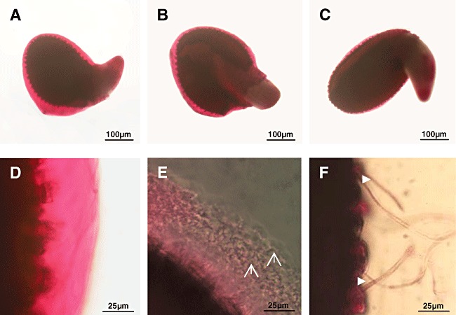
Investigations of the seed mucilage during co‐cultivation.
A–F. Seeds were placed onto agar plates alone (A and D), together with spores of S. lividans (B and E), conidia of V. dahliae (C and F) and incubated.
A–F. After 3 days, samples were stained with Ruthenium red (see Experimental procedures), and inspected by light microscopy at low (A–C) or at high magnification (D–F).Hyphae of S. lividans ( ) or of V. dahliae (
 ) are marked.