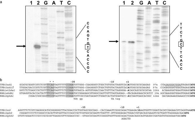
Figure 5.

Characterization of thnA1 and thnS promoter regions. A. Primer extension assays for thnA1 (left) and thnS (right) transcriptional start point mapping. Total RNA from glucose‐ (lanes 1) or from tetralin‐ (lanes 2) grown cells were used as templates for primer extension reactions. The transcriptional start point is marked with a square. B. Alignment of the thnA1 and thnS promoter sequences (coding strand) with other RHA1 promoter sequences. In bold and italic, transcriptional start points experimentally determined. Underlined, putative thnA1 ribosome binding site. Proposed −10 and −35 consensus sequences are indicated under the alignment. Putative thn boxes, important for tetralin induction, are shadowed and asterisks denote the nucleotides mutated in plasmids pMPO645 (C‐51A) and pMPO646 (G‐49T).
