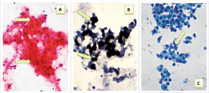
Figure 3.
Reactive ascitic fluid showing: a- Cluster of benign looking reactive mesothelial cells with low nuclear to cytoplasmic ratio (arrows) (Pap40×). b- Positive immunocytochemical expression of CAL with brown cytoplasmic and nuclear staining with strong intensity (arrows) (40×). c- Reactive mesothelial cells with negative immunocytochemical expression of CEA (arrow) (40×).

