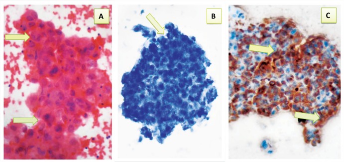
Figure 6.
Malignant pleural fluid showing: a- Cluster of malignant epithelial cells with marked pleomorphism, high nuclear to cytoplasmic ratio, hyperchromasia and prominent nucleoli (arrows) (Pap 40 ×). b- Negative immunocytochemical expression of CAL (arrow) (40×). c- Positive immunocytochemical expression of CEA with brown cytoplasmic staining with strong intensity (arrows) (40X).

