Figure 4.

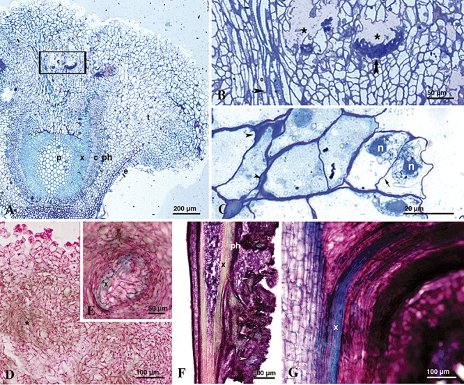
Light microscopy images of cross‐sections of knots induced by P. savastanoi pv. savastanoi NCPPB‐3335‐GFP on in vitro olive plants. A–C. Cross‐semi‐thin sections of knots induced at 35 dpi, stained with toluidine blue. (A) Pith parenchyma (p), secondary xylem (x), vascular cambium (c), secondary phloem (ph), epidermis (e) and hypertrophied parenchyma (t). Stained primary and secondary walls show dark and light blue colour respectively. (B) Detail of a hypertrophied area of the tissue showing internal cavities surrounded by collapsed host cells (asterisks), longitudinal xylem vessels running out of the stem vascular system towards the knot tissue (black arrowhead) and intensively stained cell wall accumulations (black arrow). (C) Detail of hyperplasic cells showing two nuclei (n) separated by a slim middle lamella (black arrow). Irregular thickenings of the primary cell wall surrounding parenchymatic‐like cells of outer knot layers (arrowheads). D and E. Cross‐sections of knots, collected at 28 dpi, stained with methylene blue‐picrofuchsin. (D) Groups of parenchymatous‐like cells, showing a slight blue‐green stain of the cell walls due to the formation of secondary walls during differentiation (asterisks). (E) Detail of a newly formed bundle of xylem vessels (x). F. Panoramic view of a longitudinal section of a knot, collected at 21 dpi, stained with methylene blue‐picrofuchsin. Bundles of xylem are distinguished inside the hypertrophied area (white arrowheads). G. Detail of a vascular connection between the vascular cylinder and the hypertrophied parenchymatic tissue observed in a longitudinal section of a knot collected at 21 dpi.
