
Figure 5.
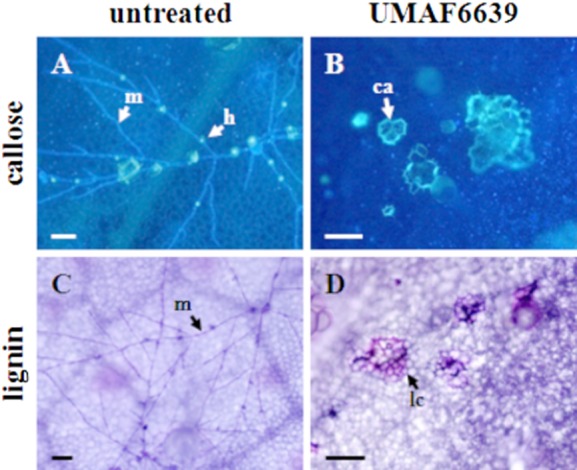
Histochemical analysis of cell wall reinforcement in leaves of melon plants bacterized with B. subtilis UMAF6639 and infected by powdery mildew. Melon plants were bacterized and inoculated with P. fusca as described in Experimental procedures. A and B. Detection of callose deposits (ca) surrounding the cells by calcofluor staining and fluorescence microscopy. Haustoria (h) can be distinguished as blue fluorescent spots along P. fusca hyphae (m). C and D. Lignin deposition analysed by toluidine staining and bright-light microscopy. Micrographs show lignified cells (lc) and P. fusca mycelia (m) both stained in violet. Pictures were taken 72 h after inoculation of the fungal pathogen. Scale bars represent 500 μm (B) and 50 μm (rest of the plates).