Figure 4.
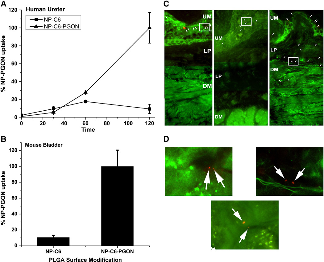
Uptake of functionalized coumarin-6 and nile-red NPs in human ureter and intravesically instilled mouse bladder. (A) Human ureter was exposed to control NPs (NP-C6) or NP-C6-PGON for 2 h and uptake of fluorescence measured. Data are expressed as % uptake of NP-C6-PGON at 120 min (n = 3 samples each from 2 ureters). (B) Fluorescence was measured in intravesically instilled mouse bladder after treatment for 2 h with NP-C6 control (n = 8) and NP-C6-PGON (n = 6). (C) Cross-sectional fluorescence microscopy images of mouse bladder intravesically instilled for 2 h with unmodified (Plain, left panel) and modified (PEG, center panel, and PGON, right panel) PLGA-NPs encapsulated with nile-red. The white arrows indicate the nile-red NPs. Multiple fields of view (at magnification, ×400) were joined to produce a continuous bladder image containing urothelium (UM), lamina propria (LP), and detrusor muscularis (DM). The scale bar represents 50 µM. (D) Plain (left), PEG (center), and PGON (right) represent enlarged areas of the original images (C) which are defined by a white square.

