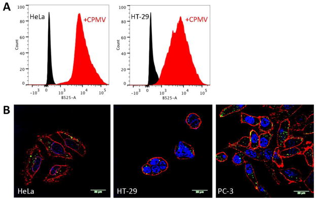
Figure 6.

A) Cell binding of A555-CPMV-PF to HeLa and HT-29 cells after 60 min exposure. For these experiments cells were collected using non-enzymatic cell dissociation buffers to avoid the natural CPMV receptor being cleaved off the cell surface; we have not achieved collection of PC-3 cells using this method. B) Confocal microscopy images of HeLa, HT-29, and PC-3 cells after incubation with A555-CPMV-PF. Red = cell membrane (WGA staining), blue = nuclei (DAPI staining), and green = CPMV (from A555 dye). The scale bar is 20 μm.
