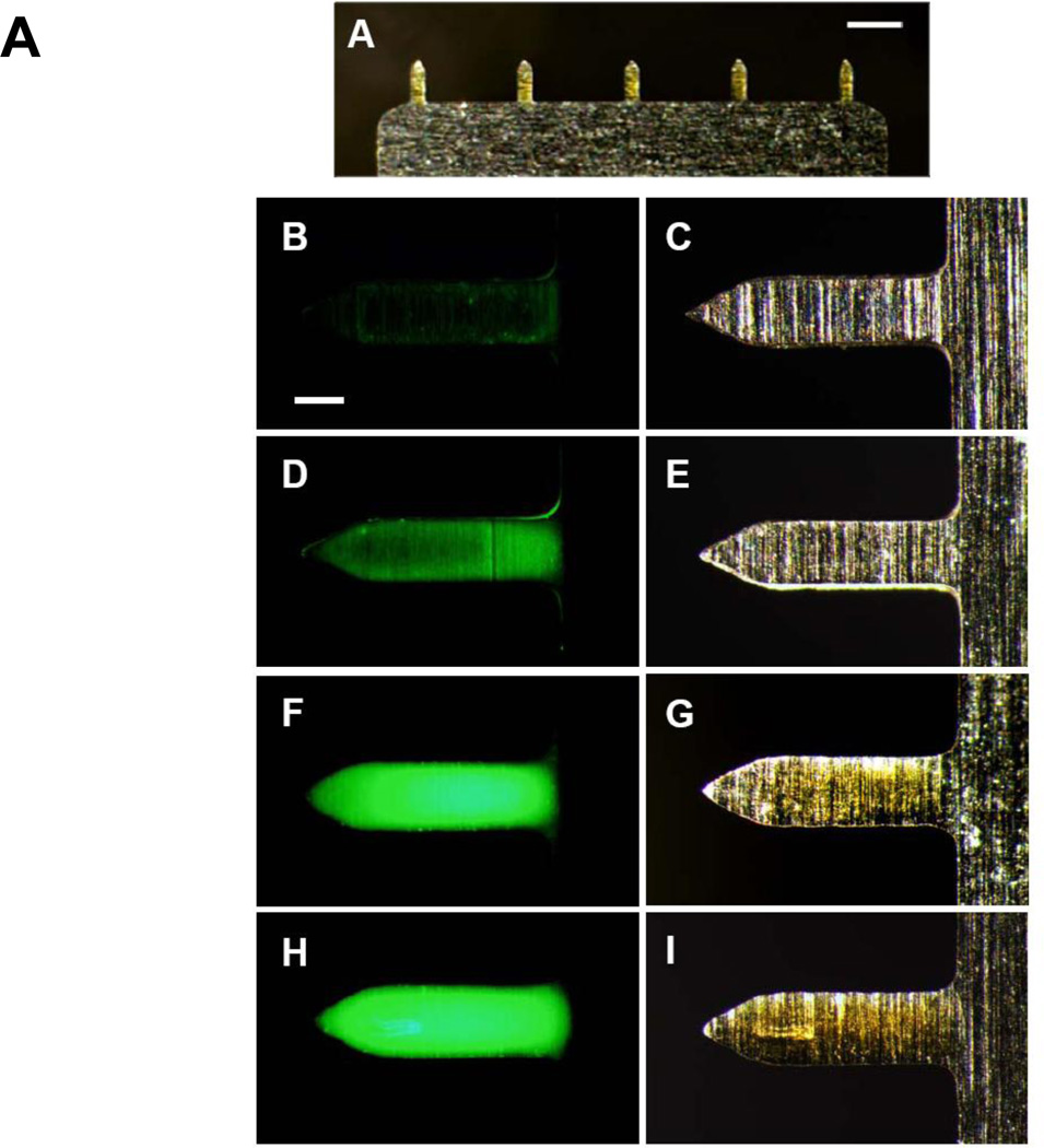
Figure 1.
Microneedles coated with inactivated virus and DNA influenza vaccines. (A) Representative array of five microneedles coated with fluorescein-conjugate BSA shown by bright-field microscopy (scale bar = 800 µm). The coating solution contained 6 mg/ml placebo DNA (B, D, F, H) Fluorescence and (C, E, G, I) bright-field microscopy images of individual microneedles coated with fluorescein-conjugate BSA coated using a coating solution containing (B, C) 2 mg/ml (D, E) 4 mg/ml (F, G) 6 mg/ml (H, I) 8 mg/ml placebo DNA (scale bar = 150 µm).

