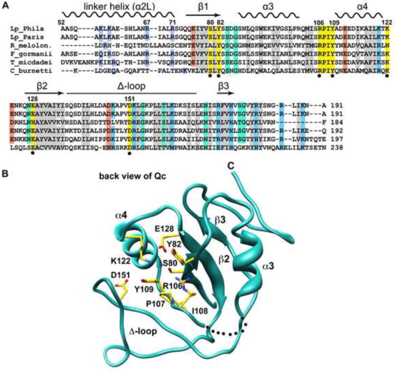
Figure 3.

Conserved residues in the Qc domain may play a role in NAD+ binding.
A. A structural alignment is shown for IcmQ sequences from 5 genera. Similar hydrophobic residues are colored grey, positively charged residues are light blue, negatively charged residues are orange, and hydrophilic residues are green. Residues that may interact with NAD+ are colored yellow and are marked with a black dot.
B. A back view of the Qc domain is shown. Conserved residues in the vicinity of the scorpion motif are labeled and their side chains are colored yellow.
See also Figure S4.
