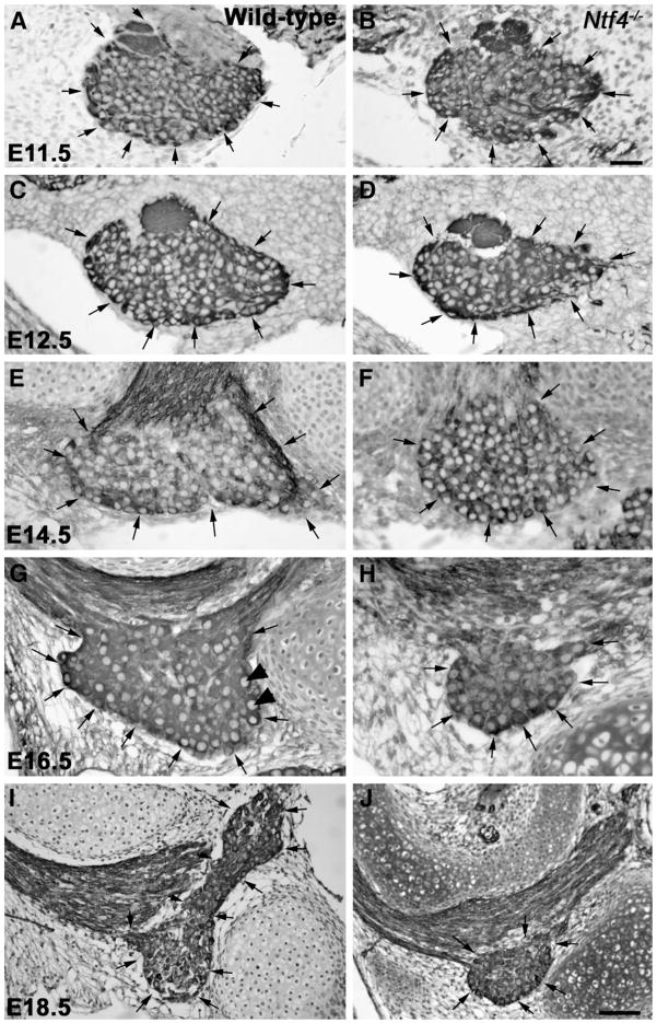
Fig. 1.
Anti-TUJ-1-labeled geniculate ganglia (arrows) from Nft4−/− mice (B, D, F, H, J) are smaller than those of wild type mice (A, C, E, G, I) beginning at E12.5. At E11.5, there is no obvious difference in the size or appearance of the geniculate ganglion between wild type (A) and Ntf4−/− mice (B). Beginning at E12.5, the Ntf4−/− geniculate ganglion (D) is noticeably smaller than the wild type (C) ganglion. These differences were also observed at E14.5 (E, F), E16.5 (G, H) and E18.5 (I, J). Note that the ganglion is much larger at E18.5 (J) compared to E16.5 (H) for wild type and Ntf4−/− mice, and, thus, imaged at a lower magnification. TUJ1 labeled neurons with a large pale nucleus were quantified as neurons (arrowheads in G). Scale bar in B=20 μm (applies to A–H); inset in H, scale bar=5 μm; J=50 μm (applies to I and J).

