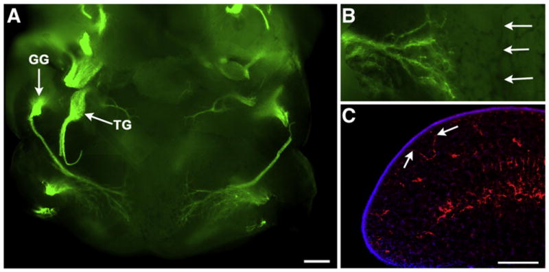
Fig. 6.

Lower jaw whole mount from an E11.5 mouse embryo labeled with anti-neurofilament (green, A, B). The geniculate ganglion (GG) and the trigeminal ganglion (TG) are brightly labeled green. Fibers from the geniculate ganglion can be seen entering the lateral edges of the tongue. At higher magnification we did not see any fibers nearing the tongue midline (B, arrows). In sections through the lateral edges of the tongue (C), some labeled fibers (anti-neurofilament, red) were seen nearing the epithelial surface (arrows, DAPI, blue). Scale bar in A=300 μm; scale bar in C=200 μm and also applies to B.
