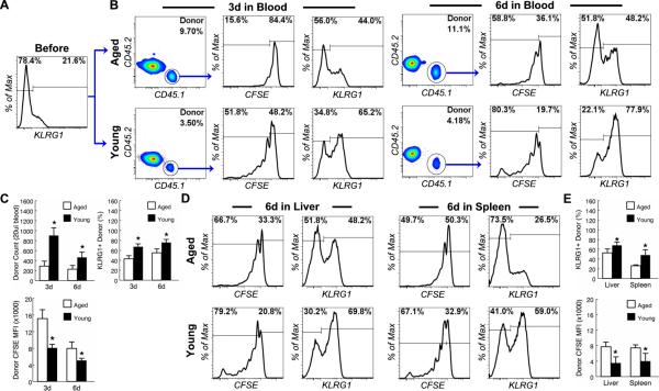
Figure 3. NK cells from young mice fail to proliferate and up-regulate KLRG1 in aged mice in response to pathogen-derived products.
NK cells from the spleen of young (3 mo.) CD45.1+CD45.2- C57BL6 mice were enriched by MACS negative selection, labeled with CFSE and transferred to young (4-6 mo.) and aged (20-22 mo.) CD45.1-CD45.2+ congenic mice. The recipients were injected i.p. with 10 ug of LPS and 50 ug of poly IC 1 day after cell transfer. (A) Donor NK cells were analyzed before transfer. Gated NK cells are shown to demonstrate the proportion of KLRG1+ cells in donor NK cells. (B) Donor NK cells from the blood were analyzed 3 and 6 day after LPS and poly IC injection. Gated donor NK cells from young and aged hosts are shown to demonstrate the proportion of KLRG1+ cells and the fluorescent intensity of CFSE labeling. (C) The mean and standard deviation of the number of donor NK cells, CFSE intensity of the donor NK cells and the proportion of KLRG1+ donor NK cells are presented. Data are representative of 2 experiments (n = 4). * indicates statistical significance between young and aged donor cells with P <0.05. (D and E) Donor NK cells from the liver and spleen of recipient mice were analyzed 6 days after LPS and poly IC injection. (D) Gated donor NK cells are shown to demonstrate the proportion of KLRG1+ cells and the fluorescent intensity of CFSE labeling. (E) The mean and standard deviation of CFSE intensity of the donor NK cells and the proportion of KLRG1+ donor NK cells are presented. Data are representative of 2 experiments (n = 4). * indicates statistical significance between young and aged donor cells with P <0.05.

