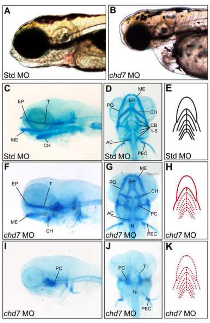
Figure 3. chd7 morphants display variable defects in craniofacial cartilage development.
(A–B) Bright-field lateral views of representative Std control morphants and chd7 morphants at 4 dpf. (C–D) Lateral and ventral views of Std morphants with wild-type craniofacial cartilage structures at 4 dpf. (F–G) Representative lateral and ventral views of the average chd7 morphant phenotype and is categorized as underdeveloped. The ceratohyal cartilages of the chd7 morphant are malformed and form a more linear shape. The five ceratobranchial cartilages were also undetectable with Alcian blue staining. (I–J) Lateral and ventral views of a severe chd7 morphant phenotype detected a highly underdeveloped neurocranium with the anterior and branchial arches absent. (E,H,K) Schematic views of the zebrafish craniofacial cartilage excluding the neurocranium. A solid red line indicates that the structure is present but malformed; while, a dashed red line indicates that the structure is absent. AC = auditory capsule, CB = ceratobranchial, CH = ceratohyal, EP = ethmoid plate, ME = Meckel’s cartilage, N = notochord, PC = parachordal, PEC = pectoral fin, PQ, palatoquadrate, T = trabecula cranii.

