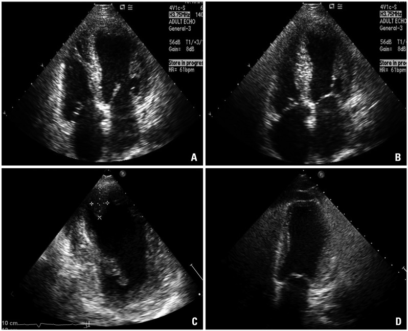
Fig. 2.
Initial echocardiography showing apical ballooning at diastole (A) and at systole (B) of apical 4 chamber view. Follow-up echocardiography showing a newly developed thrombus in the left ventricular apex 3 weeks later (C). Akinesia of the left ventricular apex was persistent but slightly improved. Follow-up echocardiography 3 months later showing persistent apical ballooning with resolution of the thrombus (D).

