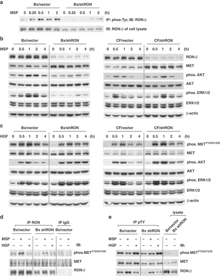
Figure 4.
Knockdown of RON enhances HGF/MET signaling. Subconfluent cells were serum starved for 16 h and stimulated with 200 ng/ml of MSP or 50 ng/ml of HGF for the indicated time periods (for 30 min in d and e). (a, b) Cells were stimulated with MSP. (a) 500 μg of total cell lysates was subjected to immunoprecipitation using anti-phospho-tyrosine (4G10) monoclonal antibody and MSP-induced phosphorylation of RON-β was detected with anti-RON-β antibody. (b) Cell lysates (40 μg) were analyzed by western blotting and phosphorylation of AKT and ERK1/2 was detected with indicated antibodies. β-Actin was used for the protein loading control. (c) Cells stimulated with HGF. Cell lysates (40 μg) were analyzed by western blotting for phosphorylated forms of MET, AKT and ERK1/2 using the indicated antibodies. β-Actin was used for the protein loading control. (d) 500 μg of cell lysates was subjected to immunoprecipitation with anti-RON-β antibody. MSP-induced phosphorylation of MET was detected by phospho-MET antibody. Co-association of MET with RON-β was detected with MET antibody. (e) 500 μg of cell lysates was subjected to immunoprecipitation with anti-phospho-tyrosine (4G10) antibody. The phosphorylation of RON-β or MET was detected with indicated antibodies.

