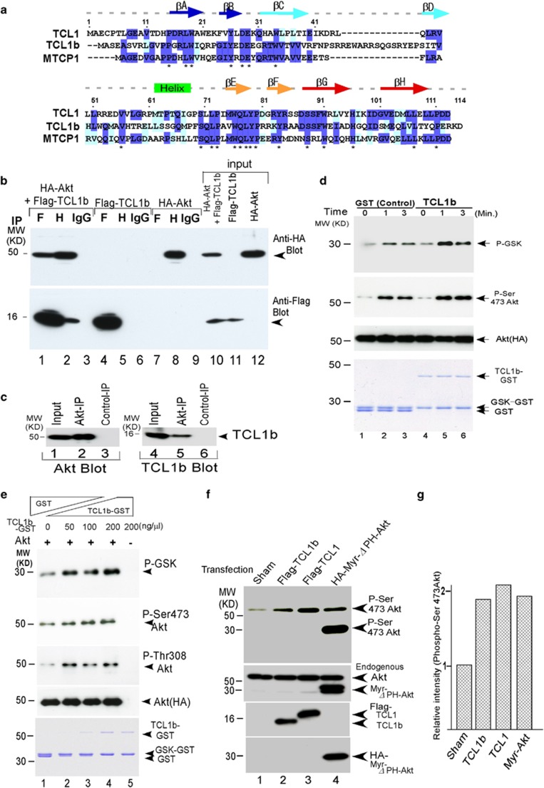
Figure 1.
TCL1b interacted with Akt and enhanced Akt kinase activity. (a) Amino-acid sequence alignment of the human TCL1 protooncogene family are shown using Clustal W (http://www.ch.embnet.org/software/ClustalW.html). (b) HA-tagged Akt2 and Flag-tagged TCL1b (as indicated) were transfected into 293T cells, and co-imunoprecipitation assays were conducted. HA-tagged Akt2 co-immunoprecipitated with Flag-tagged TCL1b in this assay. Single transfectants are shown as negative controls. There was no detectable background using control antibody. (H=anti-HA antibody; F=anti-Flag antibody). (c) In COS-7 cells, endogenous TCL1b interacted with Akt, albeit weakly, shown by using immunoprecipitation assays. (d) In in vitro kinase assays, presence of 9.6 μg of TCL1b-GST fusion proteins (0∼200 ng/μl) enhanced and promoted the kinetics of Akt-induced GSK-α phosphorylation (top panel; lanes 1–3;2866, 41852 and 36067 with control GST protein, lanes 4–6;3237, 55031 and 58123 with TCL1b). Analogously, Akt phosphorylation on Ser 473 was enhanced (second row; lanes 1-3; 18619, 26468 and 24667 with control GST protein, lanes 4–6; 14842, 55109 and 39965 with TCL1b protein). The amount of the TCL1b-GST protein or the control GST protein in the reactions is shown by coomassie brilliant blue staining (bottom row). Equal amounts of immobilized Akt were used as verified by using western blotting against HA (Third row). The levels of signal intensities were measured using NIH ImageJ. (e) The presence of 0–6.4 μg of TCL1b-GST fusion proteins in in vitro kinase assays enhanced Akt-induced GSK-α phosphorylation (top row), Akt phosphorylation of Ser473 (second row) and Thr308 (3rd row) in a dose-dependent manner. Equal amounts of immobilized Akt were used as verified by using western blotting against HA (fourth row). The amount of TCL1b-GST protein, GSK-GST and GST protein in the reactions were shown by using coomassie brilliant blue staining (bottom row). (f) Analogous to Myr-Akt (Myr-Akt (HA-Myr-ΔPH (4-129) human-Akt1 in pECE vector54), a constitutively active form of Akt, transfection of TCL1 or TCL1b into 293T cells enhanced Akt kinase phosphorylation at Ser 473. Equal amounts of Akt, each of the Flag-tagged proteins, and HA-tagged Myr-Akt were expressed as verified by using western blotting (lower panels, anti-Akt antibody, Flag antibody or HA antibody). Please note that as the PH domain is deleted in this Myr-Akt construct(HA-Myr-ΔPH (4-129) human-Akt154), the size of the band on sodium dodecyl sulfate (SDS) gel exhibited in 30 kDa in size on SDS gels. (g) Quantitation of the signals of phospho-Ser 473 Akt in panel (f) is measured and displayed as bar graph using NIH ImageJ.

