Figure 4.
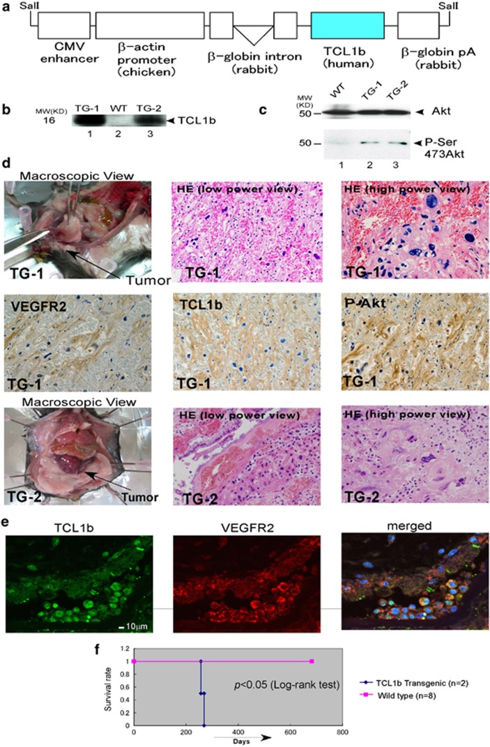
TCL1b-transgenic mice developed angiosarcoma. (a) CMV-β-actin-driven TCL1b-transgenic constructs (pUC-CAGGS-TCL1b31) were shown schematically. (b) The expressions of TCL1b of the two independent lines of transgenic mice were also confirmed by using immunoblot. (c) The levels of phospho-Akt of the muscle tissue from the two lines of transgenic mice are shown by anti-phospho-Akt (Ser473) immunoblot (lower panel) with anti-Akt immunoblot as an internal control (upper panel). (d) After 8 months of DOB, two independent lines of β-actin promoter-driven TCL1b-transgenic mice developed angiosarcoma in the intestinal submucosal tissues, which formed a huge, well-demarcated submucosal mass. Macroscopic view and HE staining (low-power view and high-power view) of the two independent lines of transgenic mice are shown (TG-1 and TG-2, top and bottom row, respectively). Microscopically, the tumor cells form irregular anastomosing vascular channels, have large nuclei and prominent nucleoli, as observed using HE staining. Immunohistochemically, the tumor cells were positively stained with anti-VEGFR2, TCL1b and phospho-Ser-473Akt antibodies (P-Akt) (middle panels). (e) TCL1b is expressed with VEGFR2, as determined by the co-staining of anti-VEGFR and anti-TCL1b antibodies and examined using confocal microscopy. (f) Kaplan–Meier curves of the transgenic lines using wild type as a control are shown. Statistically significant difference of the survival rate is observed between wild-type and TCL1b-transgenic lines (P<0.05, log-rank test).

