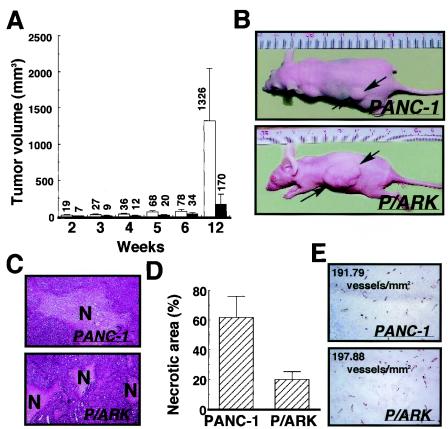
FIG. 5.
PANC-1 cells (closed bars) and P/ARK cells (open bars) were transplanted into nude mice (n = 15), and the mice were observed for 12 weeks after transplantation. (A) Tumor volume; (B) photos of tumor-bearing mice. The tumor volumes are the means of 15 data, and the bars represent the standard errors. (C) At 20 weeks after transplantation, the tumor foci formed by the PANC-1 cells and P/ARK cells were extracted and stained with hematoxylin and eosin. The necrotic area is indicated by N. (D) The necrotic area of each tumor focus was measured, the ratios of the necrotic areas are shown as the means of five data, and the bars represent the standard errors. In this study, tumor tissues were used. (E) Immunohistochemical analysis of tumors with anti-mouse CD31 antibody. The numbers on the photographs are the calculated microvessel densities.

