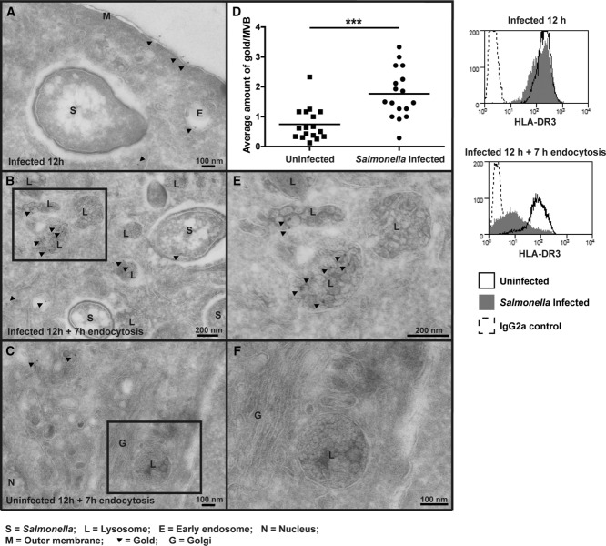
Figure 1.
MHC-II accumulates in MVBs in Salmonella-infected cells. MelJuSo were infected for 20 min with invasive GFP-S. Typhimurium (MOI 50). Cell surface MHC-II was labelled (L243) at 12 h post-infection and then cells were fixed (A) or further incubated until 20 h post-infection before fixation (B, C, E and F). Cell sections were processed for cryo-immunoelectron microscopy and HLA-DR localisation was visualised with Protein A-gold (10 nm). (D) Graph represents average amount of gold (HLA-DR)/MVB in each cell analysed. Average amount of gold/MVB was calculated for at least 15 cells per condition and comparison of distributions was assessed by unpaired two-tailed t-test. Boxed areas from (B) and (C) are magnified twofold in (E) and (F), respectively. Histograms show surface HLA-DR measured by flow cytometry in infected and uninfected MelJuSo at time points indicated. Refer to Supporting Information Fig. 1A for gating strategy. Data are representative of two independent experiments.

