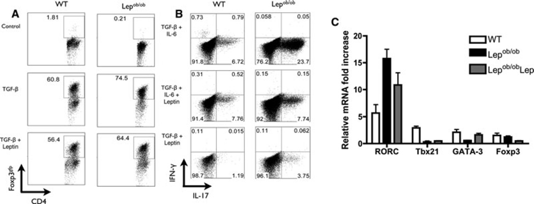
Figure 3. Tregs and Th17 cells from Lepob/ob naïve T cells are more effectively induced than those from WT naïve Lepob/ob T cells.
Naive CD4+CD62L+CD44−CD25− T cells from wild-type or Lepob/ob B6 mice were cultured for 5 days in the presence or absence of TGF-β, IL-6 and/or leptin. (A) Treg differentiation of naïve CD4+ T cells from WT and Lepob/ob mice. The induction of Foxp3+ CD4+ T cells was evaluated through the intracellular staining of Foxp3, which was then quantified in CD4+ T cells by flow cytometry. The data are representative of three independent experiments. (B) Th17 differentiation of naive CD4+ T cells from WT and Lepob/ob mice. The induction of IL17+ CD4+ T cells was evaluated through the intracellular staining of IL-17 and IFN-γ, which was then quantified by flow cytometry. The data are representative of three independent experiments. (C) The mRNA expression of RORC, Tbx21, GATA-3 and Foxp3 were analyzed after 5 days of in vitro Th17 differentiation of naïve CD4+ T cells. The total mRNA was isolated and used to prepare the complementary DNA. The mRNA levels were normalized to the GAPDH levels. Lepob/ob: CD4+ T cells from Lepob/ob cultured in the absence of leptin; Lepob/obLep: CD4+ T cells from Lepob/ob cultured in the presence of recombinant leptin; WT: CD4+ T cells from wild-type mice. The data are representative of two independent experiments. The results are shown as the mean ± SEM.

