Fig. 6.
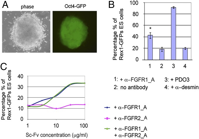
Identification of antibodies blocking FGFR1β using the ICER system. (A) Example of a colony maintaining Oct4-GFP fluorescence and ES-like morphology after 4 d of differentiation in ES-Cult/N227 following transfection with the anti-FGFR1 antibody library (magnification 20×). (B) Percentages of cells expressing Rex1-GFP after 3 d of differentiation in the presence or absence of the purified anti FGFR1β antibodies (100 μg/mL), negative (antidesmin), and positive (PDO3) controls. The bars represent the average of duplicates of a single flow cytometry experiment ± SD. (C) α-FGFR1_A and α-FGFR2_A antibodies were added alone or in combination at a range of concentrations to Rex-GFP ES cells. The percentage of Rex1-GFP+ cells from a single experiment was quantified with flow cytometry.

