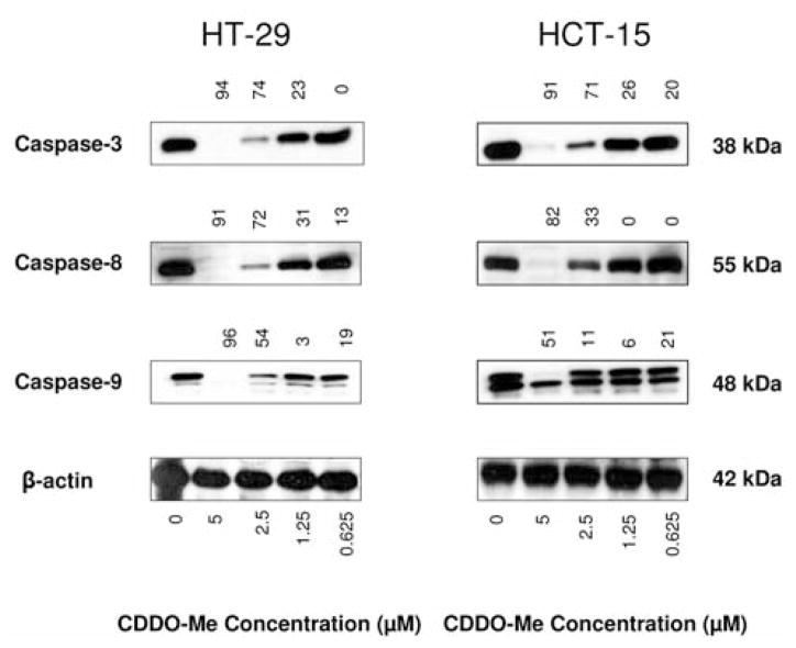
Figure 3.
Treatment with CDDO-Me cleaves procaspases-3, -8 and -9. HT-29 and HCT-15 cells were treated with CDDO-Me at concentrations of 0 to 5 μM. After incubation for 20 h, cellular lysates prepared from untreated (control) and treated cells were fractionated on 10% SDS-PAGE gel (50 μg/lane). Proteins were transferred from the gel to nitrocellulose membrane and first reacted with antibody to caspase-3, caspase-8 and caspase-9 or β-actin (loading control) followed by HRP-conjugated second antibody. Signals were visualized with enhanced chemiluminescence. Numbers above signal bands denote percentage suppression compared to control (0 CDDO-Me).

