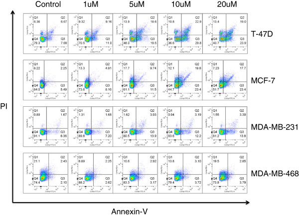
Figure 4.
Cilengitide induces apoptosis in breast cell lines. Cells were treated for 48 hours with the reported cilengitide doses and then harvested for Annexin V/PI staining and analysis by flow cytometry. T-47D cells showed the highest level of apoptotic/dead cells. MCF-7 cells showed the next highest levels of apoptotic/dead cells, with levels very similar to T-47D cells. MDA-MB-231 cells show a moderate induction of apoptosis. MDA-MB-468 cells show no apoptotic induction, which mirrors the cell detachment and proliferation assay results. Experiment was performed three times with similar results. Figures are from a representative experiment.

