
Figure 6. .
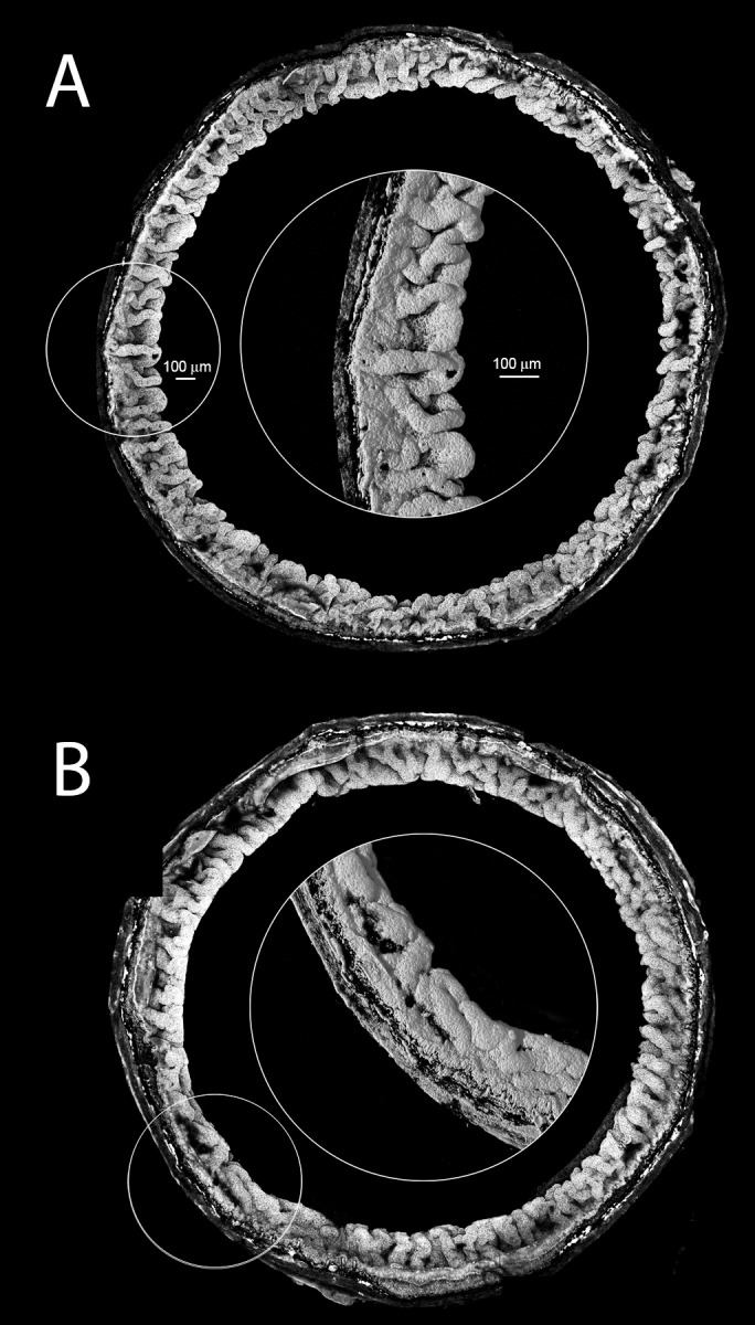
Anatomy of the ciliary body in 24-week-old WT and Fbn2−/− mice. Anatomy of intact ciliary bodies was reconstructed in three dimensions to examine the complex folding pattern of the ciliary epithelium. In WT mice (A), the ciliary epithelium is elaborately folded and the folds are predominantly radially oriented (see higher-magnification central inset). In Fbn2−/− animals (B), some regions of the ciliary body were folded normally, but in other areas (see central inset) folds were underdeveloped and circumferentially rather than radially oriented. Images are representative of four independent experiments. Scale bars: 100 μm.