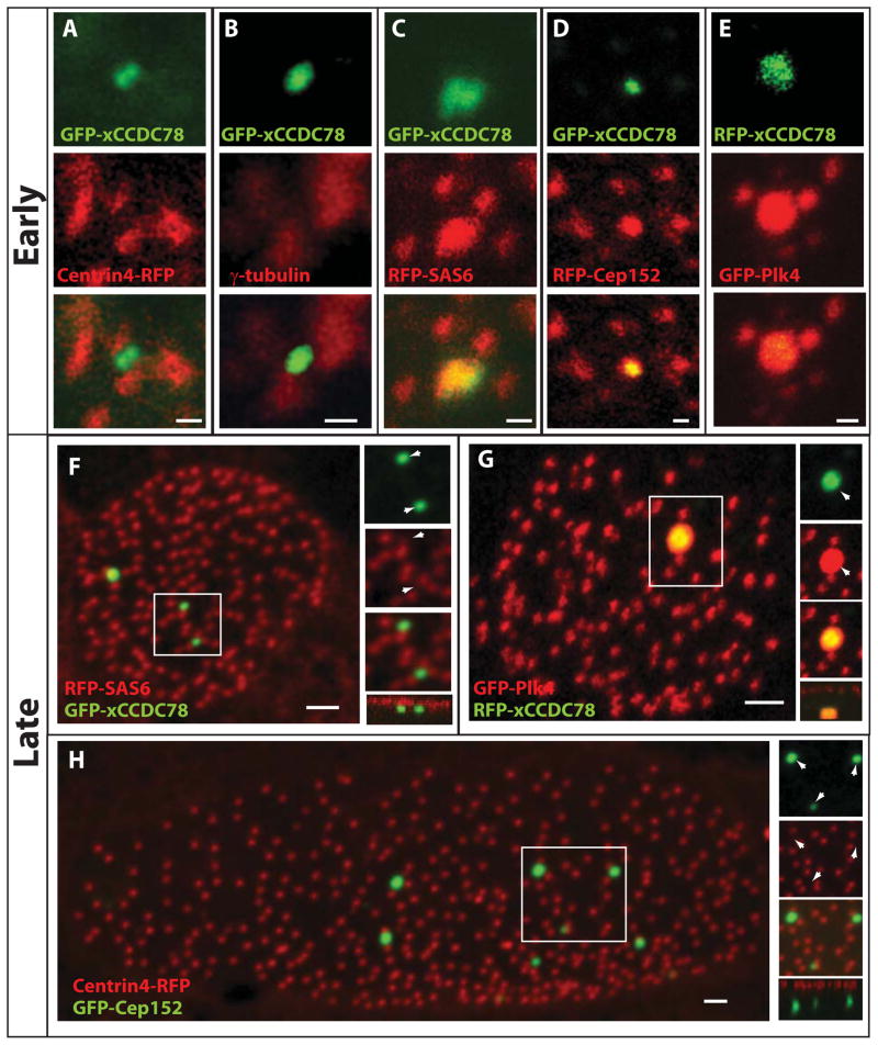
Figure 3.
Localization of key components of centriole biogenesis to the deuterosome during centriole amplification. (A–E) Localization of GFP-xCCDC78 during centriole amplification with centriolar components centrin4-RFP (A), γ-tubulin (B), RFP-SAS6 (C), RFP-Cep152 (D) and GFP-Plk4 with RFP-xCCDC78 (E) (pseudo-colored for consistency) (Scale bars, 0.5 μm). (F) Localization of GFP-xCCDC78 in mature MCC that has completed centriole biogenesis with RFP-SAS6 (F). (G) Localization of GFP-Plk4 and RFP-xCCDC78 in a mature MCC (pseudo-colored for consistency) (scale bars, 2μm). (H) Localization of RFP-Cep152 in mature MCCs to the acentriolar structure not marked by GFP-Centrin4 (scale bar, 2μm)(See also Figure S4).

