Figure 4.
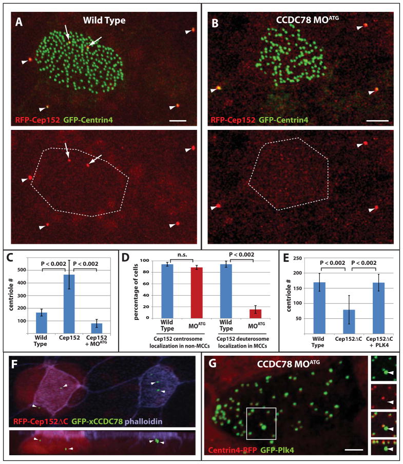
Role of Cep152 in centriole amplification. (A–B) Representative images of wild type (A) and CCDC78 morphant (B) ciliated epithelia showing co-localization of RFP-Cep152 with GFP-Centrin4 at centrosomes in non-ciliated epithelial cells (arrowheads) and the presence (A) or absence (B) of acentriolar localization in MCCs (arrows, scale bars, 5μm). (C) Quantification of centriole number in wild type embryos (n=75), embryos overexpressing Cep152 (n=42, p<0.002) and embryos overexpressing Cep152 in the presence of CCDC78 MOATG (n=86, p<0.002). Error bars, SD. (D) Percentage of cells containing RFP-Cep152 localization in the centrioles of non-ciliated epithelial cells and in the deuterosomes of MCCs in wild type (non-MCCs n=3 embryos, 335 cells, MCCs n=3 embryos, 72cells) and xCCDC78 MOATG morphant embryos (non-MCCs n=3 embryos, 396cells, MCCs n=embryos, 34cells)(Wt vs. MOATG non-MCCs, p=0.08, Wt vs. MOATG MCCs, p<0.002). Error bars, SD. (E) Quantification of centriole number in wild type cells (n=35 cells from 3 embryos), or cells expressing RFP-Cep152ΔC alone (n=34 cells from 4 embryos) or in combination with GFP-Plk4 (n=13 cells from 3 embryos). (F) Image from a mosaic embryo showing the localization of GFP-xCCDC78 in acentriolar foci in both wild type and Cep152ΔC expressing MCCs (red). (G) Image of CCDC78 morphant cell showing localization of GFP-Plk4 at both centrioles (marked with Centrin4-RFP) and at the deuterosome (no co-localization with Centrin4)(scale bars, 5μm).

