
Figure 3.
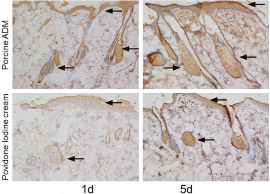
Immunohistochemistry analysis of PCNA in skin sections from two groups. Representative immunohistochemical staining images on postburn day 1 and 5. The expression of PCNA was detected in hair follicle cell nucleus and skin basal cell nucleus (brown staining, arrow). PCNA positivity was stronger in the wound tissue treated by porcine ADM than by Povidone Iodine Cream. (Original magnification ×100).