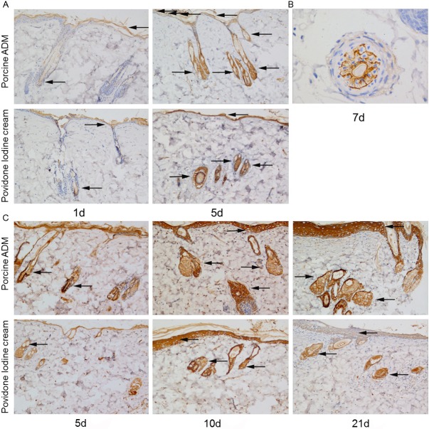
Figure 5.
Immunohistochemical staining of integrin-β1 and K19 in skin sections from two groups. A: Representative images of integrin-β1 on postburn day 1 and 5. B: Representative images of integrin-β1 on the 7th day in burn. C: Representative images of K19 on postburn day 1, 5 and 21. The expression of integrin-β1 and K19 was detected in hair follicle cells, gland cells and skin basal cells (brown staining, arrow). Integrin-β1 and K19 positivity in the epithelial cells was stronger in porcine ADM-treated wounds than in Povidone Iodine Cream-treated wounds. (Original magnification ×100 except (B), which is ×400).

