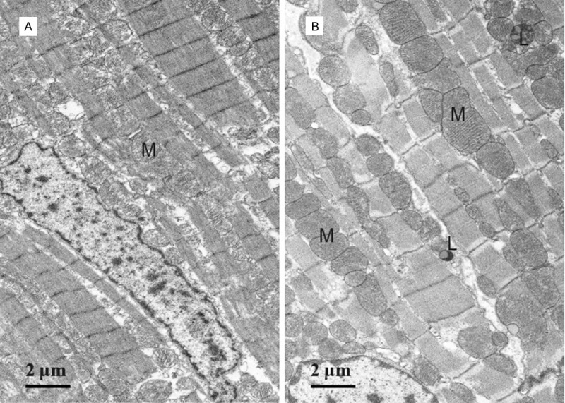
Figure 5.

Electron micrograph (TEM) of a longitudinal section of ventricular muscle. A. Control rat; B. LFN-exposed rat. Numerous enlarged mitochondria surrounding the sarcomeres are observed in the LFN-exposed rat (M). Lipofuscin granules are also observed in LFN exposed rats (L).
